
Translate this page into:
Unveiling the diagnosis of rhabdomyosarcoma on bone marrow biopsy without an obvious primary
-
Received: ,
Accepted: ,
How to cite this article: Gupta A, Kumari S, Dixit M, Jain A. Unveiling the diagnosis of rhabdomyosarcoma on bone marrow biopsy without an obvious primary. J Hematol Allied Sci 2021;1(1):41–2.
A 13-year-old girl presented with one-month history of fever, dyspnoea, pallor, multiple petechiae over bilateral lower limbs and reduced air entry in left hemithorax. CT scan chest showed multiple pleural nodules, massive left-sided effusion with multiple osteolytic lesions in the overlying ribs. Bone marrow biopsy showed infiltration by abnormal small cells with hyperchromatic nuclei forming vague nodules at places. On immunohistochemistry (IHC), these abnormal cells mimicking blasts were negative for acute leukemia markers (LCA, TdT, CD34, CD117, PAX5, CD3, MPO, CD99) and were strongly positive for Desmin, Myogenin, Myo D1, Vimentin, CD56 and negative for NSE, Synaptophysin, Chromogranin, PanCK, WT1 [Figure]. IHC findings suggested a diagnosis of infiltration of bone marrow by soft tissue sarcoma (likely to be rhabdomyosarcoma) was suggested.
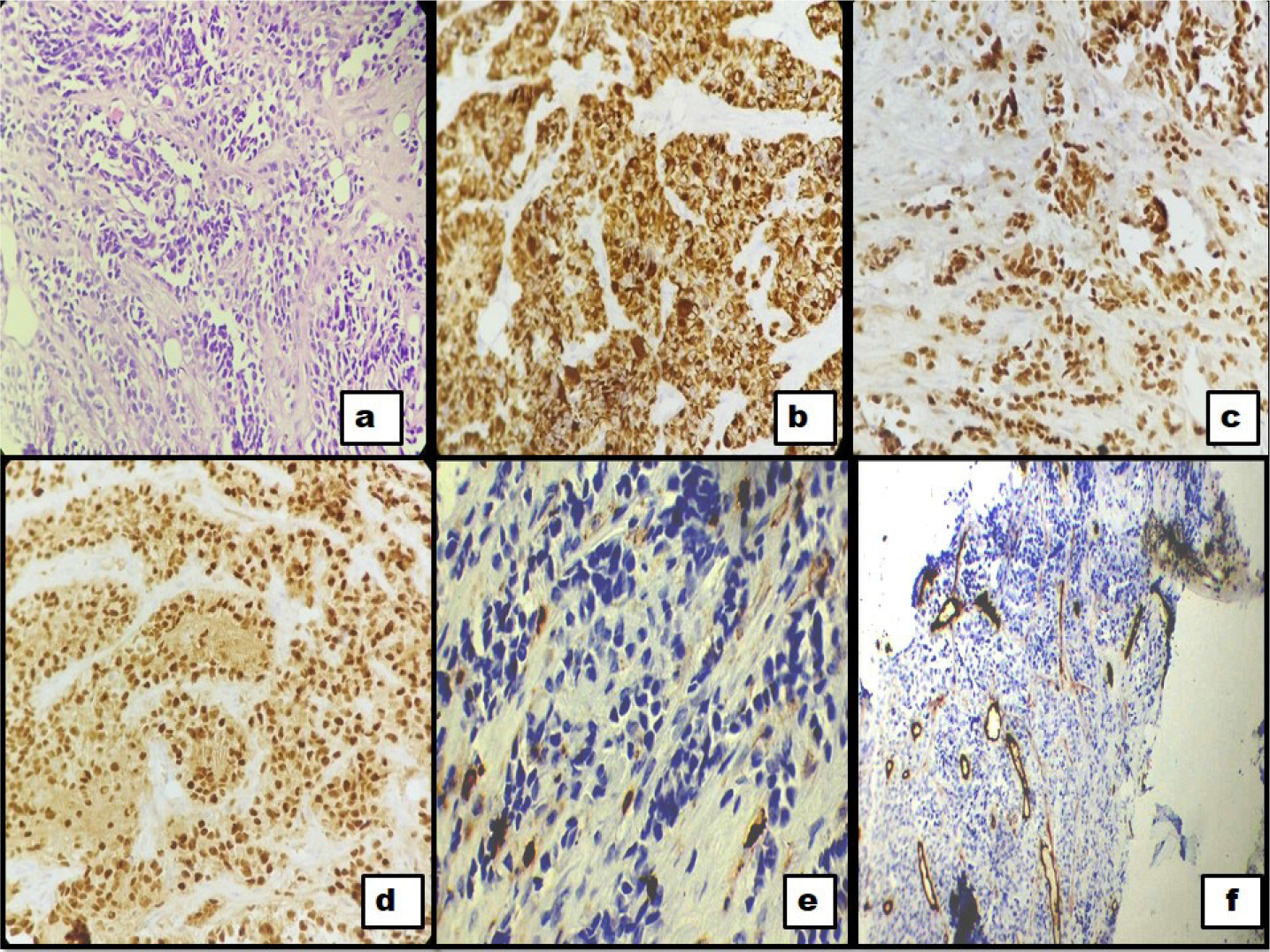
- A case of rhabdomyosarcoma with bone marrow infiltration a) H&E stained section of bone marrow biopsy (40X) with infiltration by abnormal small cells with hyperchromatic nuclei. b-d) Desmin (40X), MyoD1(40X), Myogenin (40X) shows diffuse positivity in these cells. e and f) LCA (40X) and CD34 (10X) are negative in the abnormal round cells.
Rhabdomyosarcoma is the most common soft tissue sarcoma of adolescent and childhood; however, bone marrow involvement as presenting feature without an obvious primary is an extremely rare phenomenon[1].
Declaration of patient consent
Patient’s consent not required as patients identity is not disclosed or compromised.
Financial support and sponsorship
Nil.
Conflicts of interest
There are no conflicts of interest.
References
- Bone marrow infiltration by nonhematopoetic small round cell tumors: a clinicopathological study from a tertiary care centre in South India. Indian J Med Paediatr Oncol. 2019;40:1.
- [CrossRef] [Google Scholar]


